Paralysis in pets—sudden or progressive inability to move limbs—represents a neurological emergency requiring rapid, specialized evaluation. Pet paralysis results from diverse causes affecting the nervous system, each with distinct treatment implications. Understanding paralysis causes, recognizing when surgical intervention is necessary, and knowing post-operative management requirements helps pet owners making informed decisions during pet health crises. Companion animals experiencing paralysis—whether dogs, cats, rabbits, or other species—benefit from specialized neurological expertise enabling accurate diagnosis and appropriate treatment planning.
Pet Paralysis: Causes and Pathophysiology
Diverse conditions cause paralysis.
Spinal Cord Compression and Trauma
Intervertebral disc herniation represents common paralysis cause in dogs. Herniated disc material compresses spinal cord causing varying degrees of dysfunction from pain to complete paralysis. Traumatic spinal cord injury from falls or accidents causes acute paralysis.
Neuromuscular Diseases
Myasthenia gravis, polymyositis, or other neuromuscular conditions affect muscle function causing progressive weakness and paralysis.
Peripheral Nerve Damage
Nerve injury from trauma, compression, or disease affects limb-specific function. Depending on affected nerve, specific limb patterns become paralyzed.
Central Nervous System Disease
Brain or spinal cord inflammation, tumors, degeneration, or other central disease causes progressive or acute paralysis.
Metabolic and Systemic Causes
Severe electrolyte abnormalities, toxins, or other systemic conditions rarely cause paralysis requiring careful evaluation.
Diagnostic Evaluation of Pet Paralysis
Accurate diagnosis guides treatment.
History and Physical Examination
Detailed history of symptom onset, progression, and associated signs guides diagnostic planning. Physical examination reveals paralysis pattern, location, and severity.
Neurological Examination
Detailed neurological testing reveals specific deficits localizing nervous system disease. Examination findings direct imaging location.
Advanced Imaging
MRI provides optimal spinal cord visualization revealing compression, inflammation, or degenerative changes. CT offers rapid assessment when MRI unavailable.
Laboratory and Specialized Testing
Bloodwork, CSF analysis, or electrodiagnostic testing identifies specific underlying causes.
When Surgical Intervention Is Indicated
Determining surgical necessity requires specialized expertise.
Disc Herniation and Spinal Compression
Severe disc herniation causing significant paralysis often requires surgical decompression. Surgical timing—earlier is better—dramatically influences outcomes.
Traumatic Spinal Cord Injury
While most spinal trauma cannot be surgically corrected, carefully selected cases benefit from surgical stabilization preventing additional injury.
Spinal Cord Tumors
Accessible tumors compressing spinal cord may benefit from surgical removal enabling recovery and preventing further deterioration.
Functional Improvement Potential
Surgery is considered when imaging reveals structural lesions potentially amenable to surgical correction.
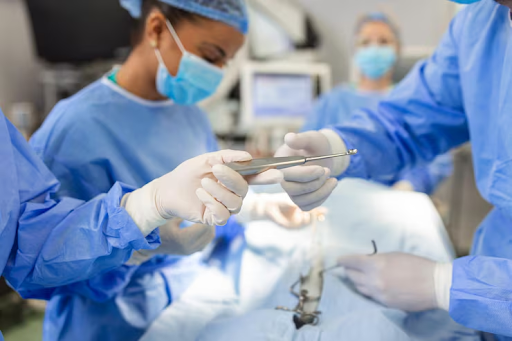
Surgical Approaches and Techniques
Various surgical techniques address spinal pathology.
Hemilaminectomy and Decompression
Surgical removal of disc material relieves spinal cord compression. Hemilaminectomy (partial vertebral arch removal) provides disc access minimizing bone removal.
Ventral Slot Procedure
Ventral slot involves removing ventral vertebral material enabling disc material removal through ventral approach. Technique selection depends on disc location.
Spinal Stabilization
Fracture stabilization through fixation prevents instability and progressive neurological deterioration.
Minimally Invasive Techniques
Emerging techniques minimize surgical trauma while achieving decompression.
Post-Operative Care and Recovery
Proper post-operative management is critical.
Wound Care and Monitoring
Incision monitoring prevents infection and complications. Regular wound assessment ensures healing.
Activity Restriction
Strict activity restriction (4-6 weeks typical) prevents destabilization and promotes healing.
Pain Management
Appropriate pain control ensures comfort supporting rehabilitation participation.
Physical Rehabilitation
Graduated rehabilitation activities support neurological recovery. Rehabilitation intensity increases as healing progresses.
Non-Surgical Management Options
Surgery isn’t always necessary or appropriate.
Conservative Management
Some paralyzed pets improve with conservative management—rest, pain control, physical therapy—without surgery.
Medical Therapy
Anti-inflammatory medication, immunosuppression (if appropriate), and supportive therapy manage select conditions.
Rehabilitation and Supportive Care
Intensive rehabilitation with mobility aids, frequent position changes, and hygiene management improve outcomes even when full recovery doesn’t occur.
Palliative Management
Some severe cases require palliative care maintaining comfort when recovery isn’t expected.
Managing Expectations and Recovery Timeline
Realistic expectations guide decision-making.
Recovery Variability
Recovery timelines vary dramatically—some pets recover rapidly; others require months. Some achieve only partial recovery.
Prognostic Factors
Severity at presentation, time to treatment, and specific underlying condition influence outcomes.
Long-Term Outcomes
Many pets achieve good quality of life with continued rehabilitation and supportive management even if complete recovery doesn’t occur.
Conclusion
Pet paralysis requires rapid specialist evaluation enabling accurate diagnosis and appropriate treatment planning. Whether surgical or conservative management is selected, specialized expertise optimizes outcomes. With proper diagnosis, appropriate treatment, and dedicated rehabilitation, many paralyzed pets achieve meaningful functional recovery or maintain good quality of life supporting their wellbeing and that of their families.