Heart conditions often develop gradually, making an accurate diagnosis essential for timely and effective cardiac care. In India, cardiologists increasingly rely on clinical data and diagnostic imaging to assess heart health with greater clarity. This data-driven approach, used by one of the best cardiologist in India, focuses on objective evidence of cardiac function rather than symptoms alone, supporting accurate diagnosis and informed clinical decision-making.
How Clinical Data Guides Cardiac Diagnosis?
Clinical data form the foundation of cardiac assessment. Cardiologists evaluate measurable indicators that reflect how the heart and related systems function over time. Instead of relying on a single report, doctors examine patterns and trends that may indicate underlying stress or imbalance.
Key clinical data points used in cardiac diagnosis include:
- Blood pressure readings recorded over time
- Cholesterol and lipid profile levels
- Blood sugar values linked to metabolic health
- Heart rate patterns and rhythm observations
- Personal and family history of heart conditions
Together, these data points help identify potential risk factors early. When patterns suggest concern, clinicians may determine whether imaging or further investigation is required. This ensures that additional tests remain appropriate and clinically relevant.
Role of Diagnostic Imaging in Heart Evaluation
Diagnostic imaging enables cardiologists to assess the heart directly rather than relying on indirect indicators. Techniques such as echocardiography, cardiac CT, and cardiac MRI provide visual information about heart structure, blood flow, and muscle movement. These images help confirm whether clinical data correspond to structural or functional abnormalities.
Imaging contributes clarity to the diagnostic process. Visualising how the heart functions at rest or under stress allows clinicians to make more confident assessments, supporting timely care planning and reducing unnecessary delays.
Echocardiography as a Core Diagnostic Tool
Echocardiography is one of the most commonly used imaging methods in cardiology. It uses sound waves to create real-time images of the heart, enabling assessment of chamber size, valve movement, and pumping function.
As a non-invasive and widely available test, echocardiography is often used early in the diagnostic process. It can help identify valve disorders, reduced heart muscle function, and structural changes. The findings may guide further investigation, ongoing monitoring, or referral for additional imaging where required.
Advanced Imaging for Detailed Cardiac Assessment
In some situations, routine tests may not provide sufficient detail. Advanced imaging techniques allow for more precise evaluation of complex or unclear findings. Cardiac CT scans provide detailed views of coronary arteries, while cardiac MRI offers insight into heart tissue health, inflammation, and scarring.
By combining advanced imaging results with clinical data, cardiologists can better understand the underlying causes of symptoms or abnormal test findings. This integrated approach supports accurate interpretation and appropriate care planning.
Integrating Data and Imaging for Informed Decisions
Accurate diagnosis depends on how effectively clinical data and imaging findings are interpreted together. Cardiologists assess imaging results alongside clinical indicators to establish relationships between symptoms, test results, and heart function. This integrated interpretation of data and imaging reflects the diagnostic approach followed by one of the best cardiologists in India, supporting accurate and well-informed clinical decisions.
This combined interpretation also helps clinicians communicate findings clearly to patients and supports consistency across care teams. When diagnosis is based on both measurable data and visual evidence, care planning becomes more precise and reliable.
Importance of Early Detection Through Monitoring
Many heart conditions progress without noticeable symptoms. Ongoing monitoring of clinical data, combined with timely imaging, allows cardiologists to detect changes early, often before complications arise.
Early detection supports improved outcomes by:
- Identifying silent changes in heart function
- Confirming concerns through targeted imaging
- Allowing timely medical review or guidance under professional supervision
- Reducing the likelihood of sudden complications
By integrating data monitoring with imaging, cardiac care increasingly focuses on early identification rather than reactive treatment.
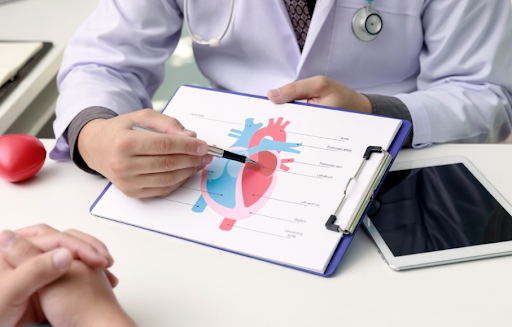
Using Diagnostic Findings to Support Patient Understanding
While technology plays a central role in diagnosis, transparent and empathetic communication remains essential. Cardiologists use data and imaging findings to support discussions with patients, often using visual explanations to clarify complex information.
Clear communication helps reduce uncertainty and supports patient understanding of their condition and care pathway. When patients feel informed, they are more likely to engage in follow-up and ongoing monitoring, making diagnosis a shared, informed process.
Conclusion
Cardiac diagnosis in India increasingly relies on the combined use of clinical data and diagnostic imaging. By integrating measurable health indicators with visual assessment, cardiologists improve diagnostic accuracy and confidence in clinical decision-making. This evidence-based approach supports early detection, informed care, and long-term patient wellbeing, while maintaining a focus on clear communication and individual needs.